


People and animals are attacked by these creatures, the infection is easily transmitted to each other through infected products, water and dirty hands.The emergence of parasites in the body will help prevent the rules of prevention and careful observance of personal hygiene, but even these measures do not guarantee one hundred percent protection.Helminants inhabiting the human body can live in various organs, differ in appearance, size, as well as the degree of harm done to a person.
Parasites in the human body
How many situations are there when a person walks around doctors for years and cannot get rid of allergies, treats asthma, drinks sugar -down drugs and all to no avail?Each of us has such acquaintances who spent huge amounts on the treatment of various diseases, and did not get any result.
Only in rare cases, when the doctor is either very smart or very responsible, does he direct such a patient to a regular analysis of feces and then ... Then parasites are found in the human body, which cause dozens of diseases, and with which no one fights in the treatment of these pathologies.
Contrary to the established opinion, worms are not necessarily “prescribed” in the intestines and can be detected by a banal analysis of feces.Many parasites feel great in the lungs, heart, muscles, even in the brain and eyes.
The well-known malaria, which was once defeated, has returned again and this is the most dangerous Parasitic disease in the opinion of experts.Malarial plasmodium lives exclusively in the blood and not every doctor will be able to recognize this disease with sufficient confidence.
How parasites fall into the human body
They fall into the human body in different ways, most often this is due to the use of infected water and food.
Grape eggs remain viability for up to 6 months and through toys, carpets, underwear and pastel linen enter the body.Askarid eggs come to us inside through poorly washed vegetables and fruits.Barbecue or homemade lard is a 95% guarantee of infection with trichinellosis.
Parasites penetrate inside us with insect bites, when bathing in freshwater reservoirs, through air, with dust, which is a carrier of eggs.
Salt fish, stroganin or caviar are the cause of infection with tape worms, the length of which reaches 12 meters and which can live in your body under 25 years.Cases of infection of infants have become more frequent in parasites in the womb.Dogs and cats, through their wet breathing, can dissipate parasite eggs at a distance of up to 5 meters.
You can get infected through dirty hands, not only their own, but also sellers, cooks, waiters, parasites travel on money and handrails of public transport.A high concentration of parasite eggs is observed in products, such as: bacon, smoked sausage, ham, sausages, pork of any shape, beef, chicken, lamb, and even chicken eggs are very often infected with them.
Epidemiologists of the whole world are trying to fight this misfortune.In the United States, for example, for verification for helminthias, 1 pork car in every thousand is destroyed.This develops in multimillion -dollar losses, but otherwise it is impossible.
There are no absolutely reliable ways of disinfecting meat, and ordinary culinary processing of larvae does not destroy.You cannot guarantee yourself the purity of your food by welding or frying meat, a huge number of parasite larvae still penetrates your body.
In which organs can a person live in parasites
Helminthic parasites are divided into two categories that correspond to the place of activity in the body of the donor:
- cramp: worms living in various gastrointestinal sections.There are about 100 varieties of intestinal parasites, and for each intestine department there are a couple of dozen species.The small intestine is ready to take ascaris, antelost, wide ribbons and other less common “brothers”.The small intestine will “share the living space” with pinworms, a dwarf chain and the rest.The medical literature describes cases when one person was infected with several types of parasites at the same time;
- Fabric:Worms localized in organs, tissues, and even in the blood.Modern medicine successfully copes with the paragonimosis (lungs), cystycchosis (brain), echinococcosis (liver) and phylairiosis (lymph vessels).Some worms of worms move through the body through the blood system and randomly attached to any organ.If many eggs have been introduced, then the whole body can be infected.
Snoes - how they look in the human body (photo of worms)
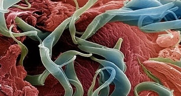
Speakers- These are one of the most common human parasites, which are a round worm (nematode).Most often, infection with pinworms occurs in children, but is also found in adults.
The pin is a white parasite, small and round.The individuals of the female individuals have dimensions: 8-13 mm length, 0.5 mm thickness, an oblong shape and a straight tail, pointed at the end.
This feature of the tail of the female parasite explains its name - “cutter”, from the word “sharp”.The male individual is much smaller: its length is 2-5 mm, the thickness is 0.2 mm, the tail is bent, unlike female pinworms.
Human invasion has the name Enterobiosis, and occurs mainly with non -compliance with personal hygiene rules (insufficient hand washing).Basically, the pinworms live in the small section of the intestine and the upper part of the large intestine, but in some cases they can also migrate to other organs and systems of organs.

The female helminth, having fallen into the human body with an oral way, and mating with the male representative of the Nematode, migrates into the thick department of the intestine, where she receives the necessary nutrients for life and maturation of eggs from undigested food residues.
After 4 weeks, the female cutter begins to migrate in the rectum at a speed of 12 cm per hour, crawls out of the anus and lays about 5000-15000 eggs in the perianal region, which after 4-6 hours are completely ripened and ready for a further life cycle.
This process can be accompanied by itching, which encourages an infected person to comb the anus, and thus contribute to the further spread of parasites, which fall into food from under the nails, into the hands of other people (especially for children who are very closely in contact with each other and not always observing personal hygiene rules).
Infection with pinworms comes from a person to a person, through the dust with the parasite eggs, objects that the patient touched.Gutty eggs can also be transferred to food with cockroaches and flies.
Also, eggs remain on linen, clothes, beds, which explains their rapid distribution.Due to the fact that the worm’s life cycle is very fleeting, and infection comes from a person to a person, it is quite difficult to get rid of parasites, since in addition to taking anthelmintic drugs, the patient’s personal belongings are necessary, and his insulation from other nematode media.
Askarides - how they look in the human body (photo of worms)
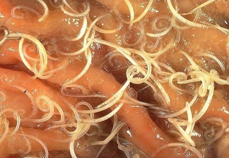
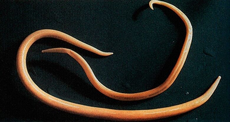
Askarida-This is a large parasite of a spindle-shaped shape of a red-yellow color, reaching an adult state of 40 cm (females) and 15-25 cm (males).Without a suction cup or other fixing devices, ascaride can independently move towards food masses.Eggs laid down by the female parasite are distinguished with feces.
Acadosis infection occurs in case of swallowing mature eggs along with water or unwashed vegetables and fruits on which there are soil particles.After the eggs penetrate the intestines, ripened larvae come out of them.
Then, introducing into the intestinal wall, they reach the heart according to blood flow, and from there they fall into the lungs.Through the pulmonary alveoli, the larva of the ascarida through the respiratory tract penetrates into the oral cavity again.
In the intestinal phase of their existence, ascarids endowed with the ability to spiral movements can penetrate even the narrowest holes.This feature of the parasite often leads to the development of quite serious complications (mechanical jaundice or pancreatitis)
After repeated swallowing, the parasite reaches the small intestine, where it develops into an adult.The worm lives for 12 months, then dies and stands out with feces.In the intestines of one owner can live both one or several hundred individuals.
Allergens secreted by ascarides can provoke severe allergic reactions.A large number of adults can cause intestinal obstruction, and worms that have penetrated the respiratory tract sometimes cause suffocation.

Vlasovsov - how they look in the human body
BlacovyvThey often live in the southern regions, since the eggs of this worm love heat.Most infections are observed in rural areas.Vlass -head eggs live in the soil.
Invasia occurs through the hands, polluted particles of the earth, poorly washed vegetables and fruits.As a result of infection, a disease occurs - trichocephalosis.Vlashev parasitizes in the intestines.This worm causes anemia, as it feeds on human blood, and severe abdominal pain.
For the diagnosis of trichocephalosis, the rectum and sigmoid intestine are examined with a special device (rectoroscopy).Thus, accumulations of parasites in the intestines are found.The treatment of invasion is long, since the sores are protected by a thick shell.
The parasite eggs stand out with feces, but they are very small, they can not always be seen even under a microscope.Only with a very strong invasion is it possible to detect eggs in the analysis of feces.In shape, they look like a barrel, have a brownish-yellow color.
On 2 sides, the eggs are holes.What do worms look like in feces?It is very difficult to find them alive in bowel movements, since the Blazoles cannot live for a long time outside the human body.Only with anthelmintic therapy can be seen in the feces of the deadworms of white color.
Hepatic bacon - how it looks in the human body
A parasite causing opisthorchiasis is a flat worm reaching a length of 7-20 mm.
In the acute phase of helminthiasis, the patient has soreness in the upper abdomen, the body temperature rises, nausea, muscle pain develops, diarrhea, and skin rashes are possible.The parasite larvae begin to develop after the eggs fall into fresh water (from the snails swallowed).Then they penetrate the body of the fish (carp, crucian carp, bream, roach).
Human infection occurs when you eat infected fish meat that has not undergone sufficient heat treatment.The larva of the hepatic bomb from the small intestine penetrates the biliary ducts and into the gall bladder, fixing there with two suction cups.
The chronic course of opisthorchiasis is manifested by symptoms of hepatitis, inflammation of the bile ducts, cholecystitis, impaired digestive tract, nervous disorders, weakness and increased fatigue.The parasite is a drive to the development of irreversible changes, and even after its expulsion, the patient does not pass chronic inflammatory processes and functional disorders.
Trichinella - what looks in the human body (photo of worms)
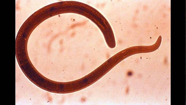
The causative agent of trichinellosis is a small round helminth that reaches 2-5 mm in length.Infection occurs when the use of poorly fried meat (pork, bear cubs, wild boar).Penetrating into the intestines, the parasite larva in 3-4 days ripens to the state of the sexually mature individual.
The life expectancy of the worm is 40 days, after which the parasite dies.Driving the intestinal wall, larvae penetrate the bloodstream and are carried in all organs of the human body, settling in the muscles.In this case, the respiratory and facial muscles, as well as muscles-ado-bends of the limbs, are most often affected.
In the first days after invasion, patients complain of abdominal pain.
Then, after about 2 weeks, the body temperature rises to 39-40 s, itchy rashes appear on the skin, muscle pain develops, and the face swells.
In this period, in the case of massive infection, there is a significant risk of death.After about a month, recovery occurs.The parasite is encapsulated in a spiral form, after which it dies within two years.

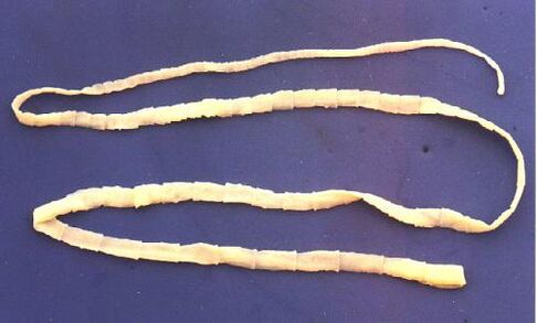
Wide tape - how it looks in the human body
This is one of the largest helminths reaching a length of 10-20 meters.The disease caused by this parasite is called dipillobotriosis.The worm development cycle begins with freshwater fish or crustaceans.
Reaching the small intestine, the parasite is attached to its wall and for 20-25 days grows to a sexually mature individual.
The larva enters the human body, which is the final owner of a wide ribbon with caviar or infected fish fillet.
Difillobotriosis proceeds against the background of disorders of the digestive tract and B12-deficiency anemia.
Echinococcus - how it looks in the body of a person
For this parasite, a person is an intermediate host.The worm parasitizes in the human body in the form of Finns.The final owner of Echinococcus is a wolf, a dog or a cat.
Infection occurs in an alimentary way in contact with animals and environmental objects, with the handful of echinococcus eggs.After getting into the intestines, oncospheres (six -black larvae) develop from them.From the intestine they penetrate the bloodstream and are carried throughout the body.
The “favorite” places of parasitization of the worm are liver and light.Settleing in these organs, the larva turns into a Finn (echinococcal cyst), which, gradually increasing in size, begins to destroy nearby fabrics.
Often, echinococcosis in the process of diagnosis is mistaken for a tumor of benign or malignant origin.In addition to mechanical exposure (squeezing organs and blood vessels), an echinococcal cyst rupture sometimes occurs.This condition can cause toxic shock or the formation of multiple new cysts.
Alveococcus - how it looks in the human body
This parasite, which is considered a variety of echinococcus, is the cause of one of the most dangerous helminthiases (alveococcosis), which is similar in severity with cirrhosis and liver cancer.Infection occurs with the penetration of oncosphere (eggs with ripened larvae) into the intestines.
This parasite, considered a variety of echinococcus, is the cause of one of the most dangerous helminthiases (alveococcosis)
There, the embryo comes out of the egg and, introducing into the intestinal walls, penetrates into the bloodstream.Further, with a blood flow, the parasite spreads through all tissues and organs of the body (most often localized in the liver).It is there that the main stage of development begins at the larvae (a multi -chamber bubble, laurelocyst) is formed).
Each chamber contains an embryo head of a parasite, which continues to gradually develop.Lavrocists are very aggressive formations that constantly grow due to increasing bubbles, as well as have the ability to germinate into the liver, like cancer metastases.
Nearby tissues due to impaired operation of blood vessels are subjected to necrotic changes.Opening nearby structures, Alveokokk forms fibrous nodes with the inclusions of multi -chamber bubbles.This condition can last for several years, in connection with which it requires compulsory surgical intervention.
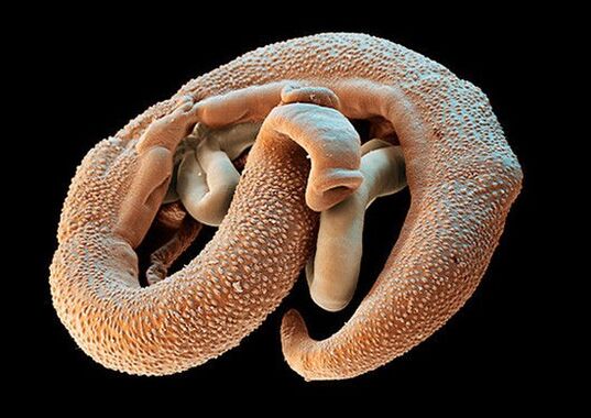
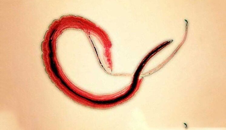
Schistosoma - what does it look like in the human body
Schistosoma - a blood -saucer, belongs to the class of trematodes, depending on the type causes various schistosomosis.This is a flat separate helminth, reaches 4-20 millimeters in length, 0.25 mm wide.The body of the schistosome is equipped with the 2nd suction cups-oral and abdominal, they are located near each other.Females of schistos is longer and thinner than males.There is a longitudinal groove on the male body, with it, he holds the female.Their eggs with a diameter of 0.1 mm, oval shape, on the surface of one of the poles are a large spike.
The human worms of the schistosomes in the role of the final owner choose people, in their organisms they parasitize in small veins of the colon, abdominal cavity, uterus, bladder.Worms feed on blood, partially absorb nutrients through the cuticle.The eggs with a schistos are transported to the intestines and bladder, where they ripen and stand out with feces or urine.In the freshwater waters, a larva - Miucidius, its intermediate host - mollusks comes out of the eggs.In the body of mollusk, metacaria develop to curials in 4-8 weeks.
Pork tinge - what does it look like in the human body
The pork tapeworm, like the bull, on the body has 4 suction cups, but in addition to this, the body of the helminth is also equipped with a double whisk of hooks.Strogil reaches two to three meters in length.The pork tapeworm has a three -doped ovary, on each side the uterus has from 7 to 12 branches.A characteristic feature of this helminth is the ability of segments to crawl out of the anus.After the exit outside, their shell becomes dry and bursts, so helminth eggs enter the external environment.The intermediate master of the tapeworm can be pigs and a person.
The main owner is a person.The intestinal parasites in humans include pork tapeworm, the helminth is located in the patient’s intestines, where he lays his eggs.Infection occurs when invasive meat is used.
Which doctor to contact with worms to contact
If infection with worms is suspected, then it is necessary to consult a doctor-infectious disease specialist, helminthologist Or a parasitologist.You can contact an infectious disease specialist regarding infection with any parasitic worms or protozoa.
You can contact a helminthologist only if infection is suspected precisely by parasitic worms (pinworms, ascarides, plumings, opisthorchiasis, etc.).
You can contact a parasitologist in cases where the infection of the protozoa is suspected - lamblia, toxoplasmes and amoebas.
In addition, if the parasite is localized not in the lumen of the intestines or stomach, but in other organs (for example, lungs, liver), then you can contact a specialist who is engaged in the diagnosis and treatment of diseases of this organ.That is, for example, with opisthorchiasis, you can also contact a gastroenterologist or hepatologist, and with echinococcosis-to a pulmonologist.